


 193 / 308

193
Micro Technology and Medical Device Technology
Micro-Macro Manipulator System
Micro-surgeries in the middle ear require the surgeon to
manipulate extremely delicate structures with fine tools,
while staying in a non-ergonomic posture for extended
periods of time. This strain on the surgeon may nega-
tively affect the outcome of the operation. Therefore, a
micro-manipulator was developed at our institute, capable
of using standard surgical instruments and controlled
via a console. In order to increase the applicability and
versatility of the micro-manipulator, a macro-manipulator
was introduced, a lightweight 6DoF robot with a reach of
0.8 m. An adapter system was developed to connect the
micro-manipulator to the tip of the robot arm. Using the
macro-manipulator drastically increases the workspace.
Furthermore, additional features were introduced, such as
a simultaneous control of both micro- and macro-manip-
ulator, hands-on-manipulation and autonomous reposi-
tioning. Additionally, a new approach to control the robot
was developed, circumventing the built-in controller and
directly accessing the communication bus. This allows for
the direct control of each individual joint by a user-written
application as well as configurations using a different
number of arm elements. The project is funded by the
German Research Foundation (Deutsche Forschungs-
gemeinschaft).
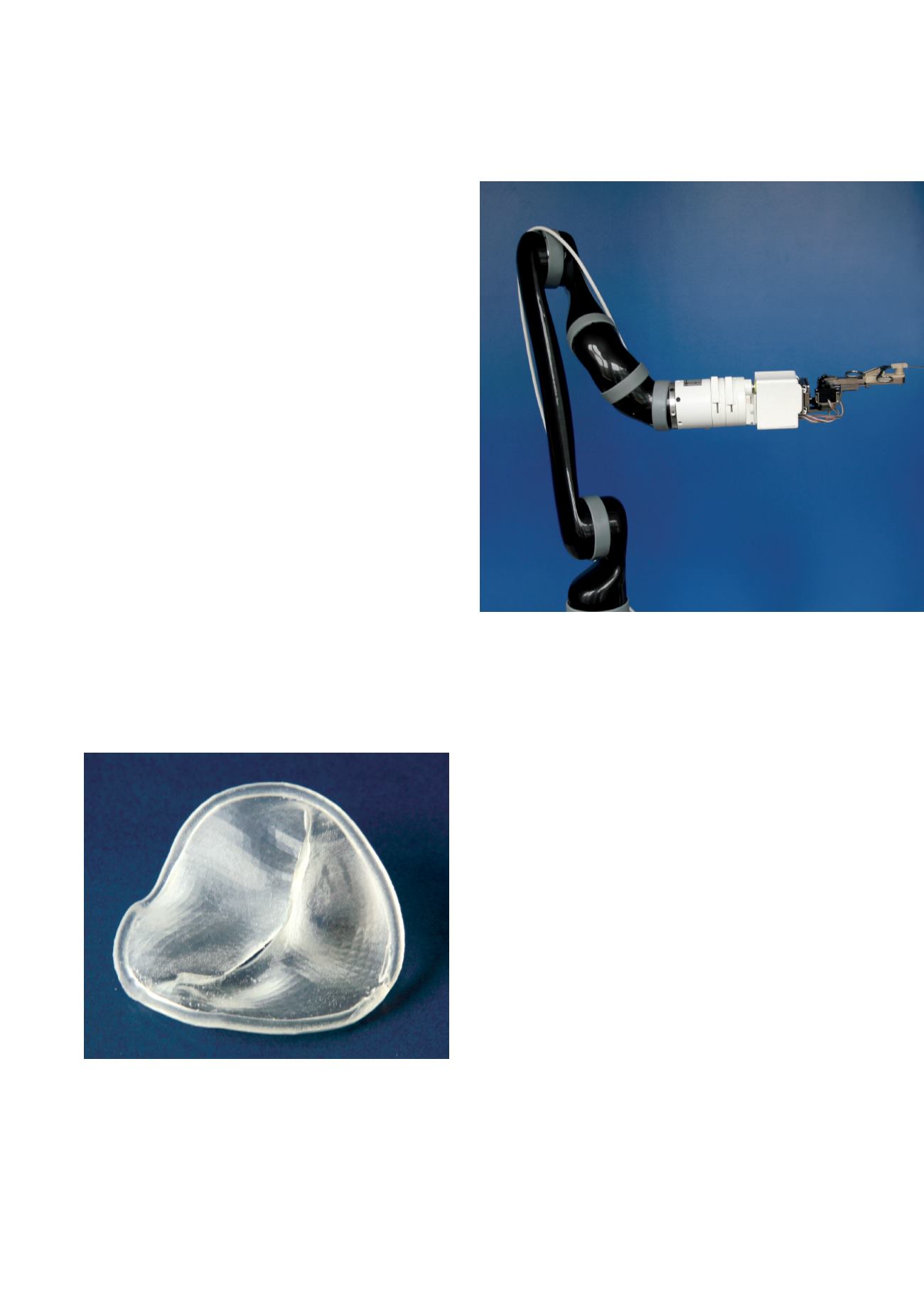
Macro-manipulator (black) with attached micro-manipulator (white)
(MiMed)
Heart valve reconstructions are complex operations. To
improve the planning of heart valve reconstructions our
institute collaborates with TOMTEC Imaging Systems
GmbH, the Klinikum rechts der Isar (Cardiovascular Imag-
ing) and the Ludwig-Maximilians-Universität München
(Cardiosurgery) in a project funded by the German Federal
Ministry of Education and Research (Support programme:
‘KMU-innovativ: Medizintechnik’, Contract number:
13GW0115A). The aim of the project is to design patient
individual 3D printed heart valve models for operation
planning and surgical templates for the realization of the
planning. From 3D ultrasound image data of patients’
hearts, the heart valves are segmented and models of the
heart valves are 3D printed. Heart surgeons can use these
models to plan the reconstruction preoperatively with con-
ventional surgical techniques. Based on the planning at
the model, a surgical template to assist the heart surgeon
with the realization of the planning during the operation is
designed.
Patient Individual 3D Printed Heart Valve Models for Operation Planning
A 3D printed model of a mitral valve (MiMed)